CELL CYCLE-CELL DIVISION
![]()
Cell Cycle
Cell cycle involves two stages
(1) Interphase or I-phase or Intermitosis or Nondividing phase or Energy phase.
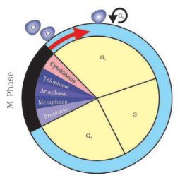
Figure: A diagrammatic view of cell cycle indicating formation of two cells from one cell
(A) G1-phase or first gap phase or first growth phase or pre synthetic phase or post mitotic phase
(B) S-phase (Synthetic phase):

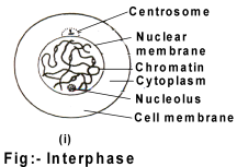
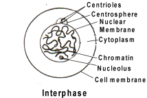
Figure: Interphase
(C) G2-phase (Second gap phase or second growth phase or postsynthetic phase or pre-mitotic
phase):
(2) M-Phase (Mitotic phase): It divides into two stages
(A) Karyokinesis (B) Cytokinesis
MITOSIS
It occurs either in diploid (animal) cells or both in diploid and haploid (plant) cells.
(A) Karyokinesis : It can be divided into four stages for sake of convinience
(I) Prophase (II) Metaphase (III) Anaphase (IV) Telophase
(I) Prophase: It is longest phase.
It involves three phases.
(a) Early prophase:
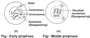
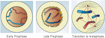
(b) Mid prophase:
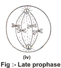
(c) Late prophase:
(II) Metaphase:
Spindle fibres consists of mostly tubulin protein, some RNA and trace amount of actin, myosin & Lipid.
(A) Continuous fibres: That connect two poles.
(B) Discontinuous fibres: They originate from a pole and do not reach at other pole.
(C) Chromosomal fibres: They connect chromosome and poles.
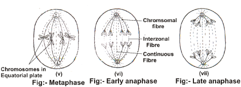
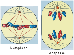
(III) Anaphase:
(IV) Telophase:

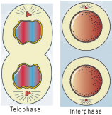
(B) Cytokinesis: It is a division of cytoplasm. It starts in late anaphase & Completed at the end of telophase.
Cytokinesis comprises two types.
(i) Cell furrow or cell cleavage method
(ii) Cell plate method
Significance of Mitosis
1. Mitosis results in two daughter cells which are gentically identical.
2. Growth and repair (cells of the upper layer of the epidermis, cells of the lining of the gut, and blood cells are being constantly replaced).
3. To restore nucleole-cytoplasmic ratio.
Key points of Mitosis
MEIOSIS

Meiosis-I: M-phase-It involves karyokinesis & cytokinesis.
Karyokinesis: It involves prophase-I, Metaphase-I, Anaphase-I, Telophase-I.
(A) Prophase- I: It is longest and complex phase. It can be differentiated into five stages.
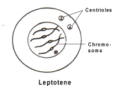
(a) Laptotene:

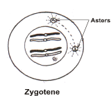
(b) Zygotene:

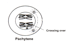
(c) Pachytene:

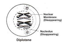
(d) Diplotene:
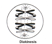
(e) Diakinesis:

(B) Metaphase-I:
(C) Anaphase-I:
(D) Telophase-I:
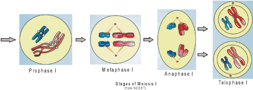
From NCERT
Interkinesis
Meiosis-II
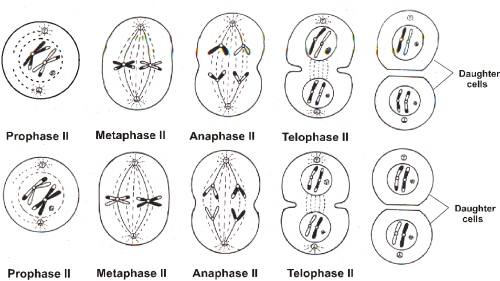
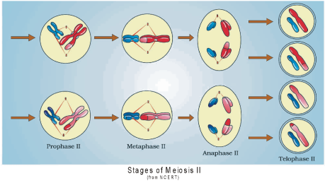
From NCERT
At the end of meiosis-II four haploid cells are formed that are genetically different.
Significance of meiosis:
(a) Variations: Variations are important for evolution.
(i) Independent assortment of chromosomes
(ii) Crossing over
(iii) Irregular disjunction
(iv) Gene mutation during replication & nicking for crossing over.
(b) Polyploidy: Failure of chromosomes to separate during anaphase-I leads to polyploidy.
(c) Maintenance of chromosome number
(d) Sexual reproduction
Key points of Meiosis:
Meiosis-I
1. Prophase-I : Devided into five stages on the basis of chromosomal behaviour.
(a) Leptotene – Bouquet stage
(b) Zygotene – Formation of synaptonemal complex, Synapsis or pairing of homologous
chromosomes and appearance of bivalent.
(c) Pachytene – Formation of recombination nodule, activity of recombinase enzymes, crossing over between non-sister chromatids of homologous chromosomes, appearance of tetrad and persistence of synaptonemal complex.
(d) Diplotene – Dissolution of synaptonemal complex, chiasmata formation due to motion of cross overed chromatids apart.
(e) Diakinesis – Terminalisation of chiasmata, disappearance of nuclear membrane, nucleolus, ER and golgi body.
2. Metaphase-I: Two metaphasic planes.
3. Anaphase-I: Separation of homologous chromosomes.
4. Telophase-I: Completion of karyokinesis-I
5. Cytokinesis-I: Two cells are formed with just half number of chromosomes as that of parent cell.
Note: Now two cells will enter into successive steps.
Interkinesis: Only deficient proteins are synthesized.
Meiosis-II
1. Prophase-II : Disappearance of nuclear membrane, nucleolus, ER and golgi body.
2. Metaphase-II: Single metaphasic plane.
3. Anaphase-II: Sister Chromatids move apart.
4. Telophase-II: Completion of karyokinesis-II.
5. Cytokinesis-II: In total four cells will be formed with half the chromosomes and DNA content that of parent cell (cell of G1 phase).
|
Difference between Mitosis and Meiosis |
|||
|
|
Mitosis |
|
Meiosis |
|
1 |
The cells undergoing mitosis may be haploid or diploid. |
1 |
The cells undergoing meiosis are always diploid. |
|
2 |
It is a single division which produces two cells. |
2 |
Meiosis is a double division. It gives rise to four cells. |
|
3 |
Subsequent mitotic divisions are similar to earlier ones. |
3 |
The two divisions of meiosis are not similar. The first one is the heterotypic or reductional while the second one is homo typic or equational like mitosis. |
|
4 |
Each chromosome replicates in the interphase before every division. |
4 |
The chromosomes replicate only once, prior to meiosis. |
|
5 |
The number of chromosomes remains the same after mitosis. |
5 |
The number of chromosomes is reduced to one half after Meiosis. |
|
Prophase |
|||
|
6 |
Prophase is of shorter duration. |
6 |
Prophase I is of longer duration while prophase II is very brief. |
|
7 |
Each chromosome has two distinct chromatids. |
7 |
Chromosomes of prophase I do not show distinct chromatids. |
|
8 |
No bouquet stage is recorded. |
8 |
Chromosomes of animals and some plants show convergence towards one side during early prophase I. It is known as bouquet stage. |
|
9 |
Pairing of chromosomes does not occur in mitosis |
9 |
Pairing or synapsis of homologous chromosomes takes place during zygotene of prophase I and continues up to metaphase-I |
|
10 |
A synaptonemal complex is absent. |
10 |
Synapsed homologous chromosome develop a synaptonemal complex. |
|
11 |
Crossing over is absent. |
11 |
Crossing over or exchange of similar segments between nonsister chromatids of homologous chromosomes usually takes place during pachytene stage. |
|
12 |
Chiasmata are absent. |
12 |
Chiasmata or visible connections between homologous chromosomes of bivalents are observed during diplotene, diakinesis |
|
Metaphase |
|||
|
13 |
Centromeres produce a single metaphasic plate. |
13 |
A double metaphasic plate is formed by centromeres in metaphase I but only one in metaphase II. |
|
14 |
Only the centromeres lie at the equator. The limbs of chromosomes are oriented in various directions. |
14 |
Limbs of the chromosomes mostly lie at the equator while the centromeres project towards the poles in metaphase I. |
|
15 |
A centromere is connected with both the spindle poles. |
15 |
A centromere is connected to one spindle pole in metaphase I but both in metaphase II. |
|
16 |
Two chromatids of a chromosome are genetically similar. |
16 |
The two chromatids of a chromosome are often genetically dissimilar due to crossing over. |
|
Anaphase |
|||
|
17 |
A centromere splits length-wise to form two centromeres in the beginning of anaphase. |
17 |
Centromeres do not divide during anaphase I but do so in anaphase II. |
|
18 |
Anaphasic chromosomes are single stranded. |
18 |
Chromosomes are double stranded in anaphase I but single stranded in anaphase II. |
|
Telophase |
|||
|
19 |
Telophase is longer and produces interphase nuclei. |
19 |
Telophase I is shorter and nuclei never enter the inter-phase. |
|
Cytokinesis |
|||
|
20 |
Cytokinesis follows every mitosis. It produces two cells. |
20 |
Cytokinesis often does not occur after the first or reductional division. It is then simultaneous after second division to result in four new cells. |
Table: Displays the various parameters changing during cell cycle (Human somatic cell with 2C DNA content for 46 chromosomes)
|
S.No. |
Phase of Cell cycle |
DNA content |
No. of chromosomes1 |
No. of Chromatids2 |
No. of Centromeres3 |
|
|
1. |
Interphase |
G1 |
2C |
46 |
46 |
46 |
|
S |
Early – 2C Late – 4C |
Early – 46 Late – 46 |
Early – 46 Late – 92 |
Early – 46 Late – 46 |
||
|
G2 |
4C |
46 |
92 |
46 |
||
|
2. |
Mitosis |
Prophase |
4C |
46 |
92 |
46 |
|
Metaphase |
4C |
46 |
92 |
46 |
||
|
Anaphase |
4C |
92 |
92 |
92 |
||
|
Telophase |
4C |
92 |
92 |
92 |
||
|
3. |
Cytokinesis – Per cell |
2C |
46 |
46 |
46 |
|
1. In interphase, there are no chromosomes as the genetic material is present in the form of chromatin material.
2. In interphase, there are no chromatids as the genetic material is present in the form of chromatin material.
3. In interphase as genetic material is not packaged in the form of chromosomes so, as such centromeres are also not defined.
Note: In all (1, 2 and 3) we have genetic material for that many chromosomes, chromatids and centromeres which will appear later in prophase.
Table: Displays the various parameters changing during cell cycle (Human germ cell with 2C DNA content for 46 chromosomes)
|
S.No. |
Phase of Cell cycle |
DNA content |
No. of chromosomes4 |
No. of Chromatids5 |
No. of Centromeres6 |
|
|
1. |
Interphase |
G1 |
2C |
46 |
46 |
46 |
|
S |
Early – 2C Late – 4C |
Early – 46 Late – 46 |
Early – 46 Late – 92 |
Early – 46 Late – 46 |
||
|
G2 |
4C |
46 |
92 |
46 |
||
|
2. |
Meiosis-I |
Prophase-I |
4C |
46 |
92 |
46 |
|
Metaphase-I |
4C |
46 |
92 |
46 |
||
|
Anaphase-I |
4C |
46 |
92 |
46 |
||
|
Telophase-I |
4C |
46 |
92 |
46 |
||
|
3. |
Cytokinesis-I Per cell |
2C |
23 |
46 |
23 |
|
|
4. |
Interkinesis Per cell |
2C |
23 |
46 |
23 |
|
|
5. |
Meiosis-II |
Prophase-II |
2C |
23 |
46 |
23 |
|
|
|
Metaphase-II |
2C |
23 |
46 |
23 |
|
|
|
Anaphase-II |
2C |
46 |
46 |
46 |
|
|
|
Telophase-II |
2C |
46 |
46 |
46 |
|
6. |
Cytokinesis-II Per cell |
C |
23 |
23 |
23 |
|
4. In interphase, there are no chromosomes as the genetic material is present in the form of chromatin material.
5. In interphase, there are no chromatids as the genetic material is present in the form of chromatin material.
6. In interphase as genetic material is not packaged in the form of chromosomes so, as such centromeres are also not defined.
Note: In all (4, 5 and 6) we have genetic material for that many chromosomes, chromatids and centromeres which will appear later in prophase.
Resonate the Concept
(1) Rudolf Virchow stated that new cell develops from the division of pre-existing cell.
(2) Strasburger stated that new nucleus arises from pre-existing nucleus. Strasburger firstly observed mitosis in plant cell.
(3) Van Beneden and Flemming discovered mitosis in animal cell. The term ‘Mitosis’ coined by Flemming.
(4) The term meiosis coined by Farmer and moore.
(5) Cyclin protein: Cyclin Dependent protein Kinases (CDK) regulate the cell cycle.
(6) Prometaphase: Some scientists considered prometaphase stage between prophase and metaphase. In this stage aster formation is completed. Formation of spindle fibres is also completed. Nuclear membrane and nucleolus are completely disappeared.
(7) Eumitosis: Formation of Extranuclear spindle and degeneration of nuclear membrane involve in it.
(8) Premitosis: In some protists, fungi and algae, formation of intranuclear spindle takes place. Nuclear membrane exists.
(9) Synapsis involves three types.
(i) Procentric: It starts from centromere and proceeds towards terminal parts.
(ii) Proterminal: It begins from terminal part.
(iii) Inter mediate type: It can begin at any point of chromosomes.
(10) Amitosis: It was discoverd by Remak. In this type, nucleus elongates& constricted in the middle and divide to form two daughter nuclei. Spindle formation is absent e.g. meganucleus of Parmaecium, cells of endosperm.
(11) Brachymeiosis: It is found in ascus bearing fungi that includes two reduction divisions and one equational division which reduce the chromosome number from tetraploid to haploid stage.
(12) Types of Meiosis:
(i) Zygotic or Initial meiosis: It occurs during zygote or zygospore germination e.g. Ulothrix, Spirogyra and Chlamydomonas.
(ii) Sporic or Intermediate meiosis: Meiosis occurs at the time of microspore or megaspore formation e.g. Bryophytes, Pteridophytes, Gymnosperms and Angiosperms.
(iii) Gametic meiosis or Terminal meiosis: It occurs at the time of gamete formation e.g. Animals
(13) Regulation of cell cycle
There are two key classes of regulatory molecules:
The cell cycle is based on three main check points.
Phase G1 – DNA integrity and cell size.
Phase S – DNA damage and duplication.
Phase M – Attachment of kinetochore and a spindle fibre.
The key role of checkpoint proteins is to detect DNA damage and send a signal to delay cell cycle until the damage chromosomes are repaired.