G.N. RAMACHANDRAN, an outstanding figure in the field of protein structure, wasthefounder ofthe‘Madrasschool’ofconformationalanalysis of biopolymers. His discovery of the triple helical structure of collagen published in Nature in 1954 and his analysis of the allowed conformationsof proteins through the use of the ‘Ramachandran plot’ rank among the most outstanding contributions in structural biology. He was born on October 8, 1922, in a small town, not far from Cochin on the southwestern coast of India. His father was a professor of mathematics at a local college andthus hadconsiderable influenceinshapingRamachandran’s interest in mathematics. After completing his school years, Ramachandran graduated in 1942 as the topranking student in the B.Sc. (Honors) Physics course of theUniversityofMadras.HereceivedaPh.D.from CambridgeUniversityin 1949. While at Cambridge, Ramachandran met Linus Pauling and was deeply influenced by his publications on models of the -helix and -sheet structures that directed his attention to solving the structure of collagen. He passed away at the age of 78, on April 7, 2001.
Theorganisms,whicharecomposedofasinglecell,arecalledasunicellularorganismswhilethe organisms, which are made up of multiple cells, are called as multicellular organisms.
Cell is a basic unit of life and it is considered as structural and functional unit of an organism. Robert Hooke (1665) discovered cell. He first observed the cell in a piece of dead cork cells. He described cell in his book “Micrographia”.
(8)New cell arises from pre existing cells “Omnis cellula-e cellula”. It is called cell lineage theory. This concept was given by Rudolf Virchow (1855). The final shape to cell theory was given by Rudolf Virchow.
Types of cells
(1)Undifferentiated cells – Also called stem cells. They are unspecialized and usually possess powerof division.e.g.-Rootandshootapices,skincells,germinalepithelium,bonemarrow, zygote etc.
(3)Dedifferentiated cells – Actually they are specialized cells but lose their specialization and induce division. It helps in healing of wounds, regeneration in animals or vegetative propagation in plants, cell culture experiments e.g. cork cambium.
.
Cell – An open system:
An open system is one which is separated from its surroundings by a boundary that allows transfer of material in and out of the cells.
Cellisanopensystembecauseitreceivesanumberofmaterialsandenergyfromoutsideand liberates energy as heat.
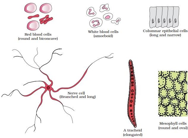
Cellsvarygreatlyintheirshape.Theymaybedisc-like,polygonal,columnar,cuboid,threadlike,or even irregular. The shape of the cell may vary with the function they perform.
Mesokaryotic cell: Histone protein absent but nucleus with nuclear membrane present. Chromosomes are condensed and visible even in interphase. e.g. Dinoflagellates.
.
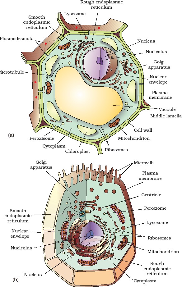
DifferencesbetweenPlantandAnimalCells
S.No.
PlantCell
S.No.
AnimalCell
1
Aplantcellhasrigidwallontheoutside.
1
Acellwallisabsent.
2
Plastidsarefoundinplantcells
2
Plastidsareusuallyabsent.
3
Amaturecellhasalargecentralvacuole.
3
Ananimalcellmayhavemanysmall vacuoles.
4
Nucleusliesononesideintheperipheral cytoplasm due to central vacuole.
4
Nucleususuallyliesinthecentre.
5
Centriolesareusuallyabsent.
5
Centriolesarefoundinanimalcells.
6
Spindleapparatusformedduringnuclear division is anastral.
6
Spindleisamphiastral.
7
Golgiapparatusconsistsofnumberof distinct and unconnected units called dictyosomes.
7
Golgiapparatusiseitherlocalisedordiffused and consists of a well connected single complex.
8
Reservefoodisgenerallystarchand fat.
8
Reservefoodisusuallyglycogenandfat.
9
Adjacentcellsmaybeconnectedthrough plasmodesmata.
9
Adjacentcellsareconnectedthrough a number of cell junctions.
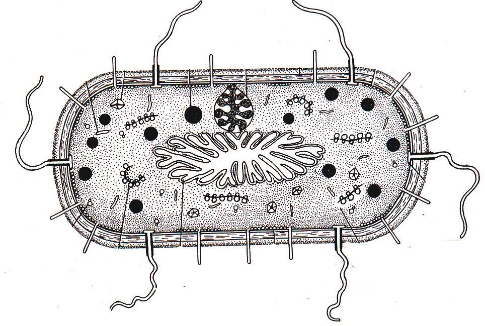
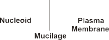
Prokaryotic cell – Cell that bears naked genetic material i.e. nucleus is without envelop is known as prokaryotic cell. This group is represented by Bacteria, Blue Green Algae (Cyanobacteria), Mycoplasma.
.
They have following characters:
(i)Like eukaryotic cells, they are different in shape and size but smaller than eukaryotes and divide rapidly.
(iv)Beside genomic DNA, small circular DNA is also present in many bacteria called plasmid which make them antibiotic resistant, regulates some phenotypes and also responsible for bacterial transformation.
(v)Most prokaryotic cells mainly the bacterial cell has envelope consists of three layers, which are tightly bound outer glycocalyx, middle cell wall and innermost cell membrane.
(vi)Although each layer of the envelope performs distinct function, they act together as a single protective unit.
(vii)If these envelopes are stained by Gram stain then they are called Gram positive bacteria while other those don’t have are called Gram negative bacteria.
(viii)Glycocalyx, a polysaccharide envelope forms either loose sheath slime layer or thick and tough structure capsule.
(xiii)Essential infoldings of Plasma membrane towards cytoplasm are called Mesosomes.They canbe in form of the Vesicles Tubules and Lamellae.
(xiv)Thesehelp in:
(a) Cell wall formation (b) DNA replication and distribution to daughter cells (c) Respiration (analogous to mitochondria) (d) secretion of processes (to increase the surface area of the plasma membrane and enzymatic content).
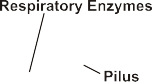
(xvii)Besides flagella, Pili and Fimbriae are also surface structures of the bacteria but do not play arole in motility. The pili are elongated tubular structures made of a special protein pilin. The fimbriae are small bristle like fibres sprouting out of the cell that provides attachment to substratum or host tissue.
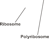
(xviii)In prokaryotes, ribosomes are associated with the plasma membrane of the cell. They are about 15 nm by 20 nm in size.
(xix)70S type of ribosomes are found in prokaryotic cells. Its two subunits are 50S and 30S. Several ribosomes are joined with mRNA to form polysome or polyribosome for efficient conduction of protein synthesis.
Cell wall not only gives shape to the cell and protects the cell from mechanical damage and infection, it alsohelps incell-to-cellinteractionandprovidesbarrier toundesirablemacromolecules. Algae have cell wall, made of cellulose, galactans, mannans and minerals like calcium carbonate.
In plant cell it is usually composed of cellulose, hemicellulose, pectins and proteins but inbacteria and BGA it is composed of peptidoglycan and DAPA. In fungi it consists of chitin. It is absent in Animals, Mycoplasma.
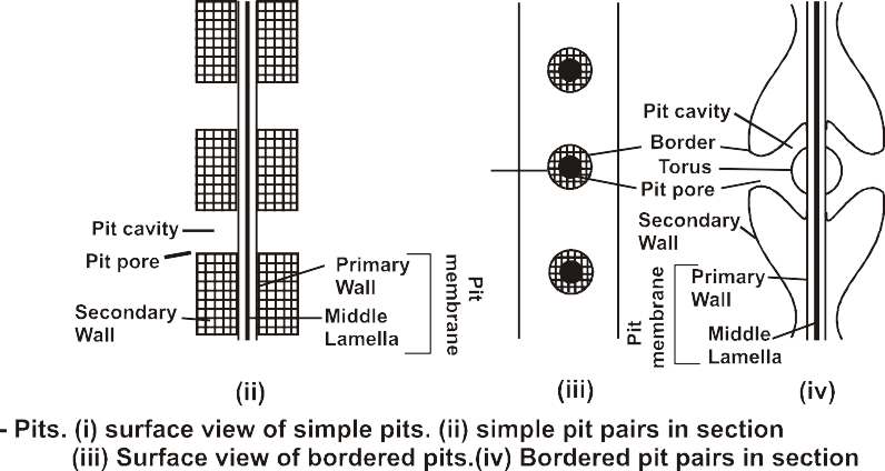
•These pits are found abundantly in tracheids of gymnosperms (have maximum number of bordered pits) and in vessels of angiosperms.
(iv)Tertiary wall:
Sometimes innermost layer of the secondary wall is distinct both chemically as well as in staining properties due to the presence of xylans. It is called tertiary wall Eg. Tension wood in gymnosperms.
.
Important point on cell wall
(1)Plasmodesmata:Thecytoplasmicbridgesbetweenadjacentplantcellsarecalled plasmodesmata. They contain E.R. tubules called Desmotubules.
(2)Expansin: It is special protein that takes part in growth of cell wall by loosing cellulose microfibrilandaddition of new cell wall materialin the space.
(4)CellCoat:Inmanyanimalsandprotistansdistinctlayerofglycocalyx isfoundintheoutersurfaceof cells. It is fibrous and composed of oligosaccharides. It helps in cell recognition, protection etc.
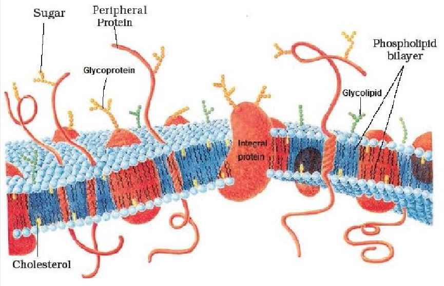
•Thedetailedstructureofthemembranewasstudiedonlyaftertheadventoftheelectron microscope in the 1950s.
•Cellmembranescanbeobservedinelectronmicroscope.Itappearstrilaminarortripartitelayer under electron microscope.
•Meanwhile, chemical studies on the cell membrane, especially in human red blood cells (RBCs),enabled the scientists to deduce the possible structure of plasma membrane.
•The ratio of protein and lipid in plasma membrane varies e.g. inhuman beings, the membrane of the erythrocyte has approximately 52 per cent protein and 40 per cent lipids.
•The plasma membrane is asymmetric due to oligosaccharides which form glycolipids and glycoprotein alongwith lipids and proteins respectively.
•Bothglycolipidsandglycoproteinsformglycocalyx.
•Oligosaccharide part in glycocalyx acts as recognition centre, site for attachment and provides antigen specificitytocellmembranes,blood grouping, immuneresponseand matching of tissues in transplantation of organs.
.
.
.
.
.
.
.
.
.
.
.
.
.
.
.
.
.
.
.
Figure:Fluidmosaicmodelofplasmamembrane
Special points of cell membrane
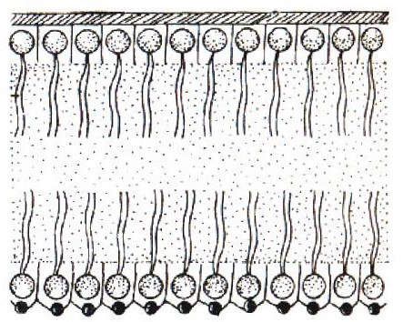
(i)Phospholipid also show exchange of molecule from one monolayer to the monolayer of other side it is called as flip-flop movement.
(vi)Neutral solutes may move across the membrane by the process of simple diffusion along the concentration gradient, i.e., from higher concentration to the lower.
(vii)Water may also move across this membrane from higher to lower concentration. Movement of water by diffusion is called osmosis.
(viii)Thepolarmoleculescannotpassthrough the nonpolar lipid bilayer, theyrequirea carrierprotein of the membrane to facilitate their transport across the membrane called as facilitated diffusion.
(ix)Some ions or molecules are transported across the membrane against their concentration gradient, i.e., from lower to the higher concentration. Such a transport is an energy dependent process, in which ATP is utilised and is called active transport, e.g., Na+/K+ Pump.
(II)Activetransport:Inthismethod,movementofsubstancesoccuragainsttheirconcentration gradient by consuming ATP. It can be done by Na+– K+exchange pump.
(III)Bulktransport:Ittakeplacebytwomethods.
.
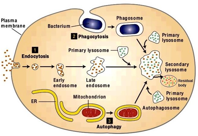
(i)Endocytosis:Theinwardtransportofmaterialbymeansofcarriervesiclesiscalled endocytosis. It includes two types.
(a)Pinocytosis or Potocytosis (Cell drinking): Intake of fluid substances by plasmalemmain the form of vesicles (Pinosome) is called pinocytosis.
(b)Phagocytosis (Cell eating):Intakeofsolidfood substancesbyplasmalemmaintheform of vesicles (Phagosome) is called phagocytosis.
(ii)Exocytosis(Cellvomittingoremiocytosis):Itisreverseofendocytosisinwhichwaste materials are removed from the cell. It involves reverse pinocytosis.
.
Cytoplasm
It lies between the nucleus and cell membrane.
In both prokaryotic and eukaryotic cells, a semi fluid matrix called as cytoplasm occupies the volume of the cell.
Cytoplasm is the main arena of cellular activities in both plant cells and animal cells. Various chemical reactions occur in cytoplasm to keep the cell in living state.
•Calcium carbonate crystals (cystolith) found in epidermal cells of momordica, hypodermal leaf cells of Banyan.
•Calcium oxalate occurs in the form of powdery mass (crystal sand) in atropa, star shaped sphaerophidein Colocasia, Begonia, Chenopodium prismatic crystals in dry scales of Onion, needle shaped raphidesin lemna, Eichhornia.
(ii)Chromoplasts: They are colouredplastids those have fat soluble carotenoids like carotene, Xanthophyll and others are present. e.g. carotene in carrot, lycopene in tomato and chillies.
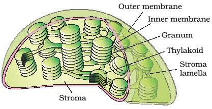
•The stroma of the chloroplast contains enzymes required for the synthesis of carbohydrates andproteins.
•It also contains small, doublestranded circular DNA molecules called cp-DNA or plastidome. and ribosomes.
•Theribosomesofthechloroplastsaresmaller(70S)thanthecytoplasmicribosomes(80S).Chlorophyll pigments are present in the thylakoids.
.
(iii)Lamellar system:
•Anumberof organisedflattened doublemembraneboundsacscalledthethylakoids,arepresentin the stroma.
•Thylakoids (2-100) are arranged in stacks like the piles of coins called grana (singular: granum) or the intergranal thylakoids. Each chloroplast has 40–60 grana.
•Intergranalthylakoids:flatmembranoustubulescalledthestromalamellaeorfretlamellae connecting the thylakoids of the different grana.
•They have originated from the symbiosis of a prokaryotic organism (aerobic bacteria) with a hostcell that was anaerobic and derived its energy only from glycolysis (Endosymbiotic hypothesis).
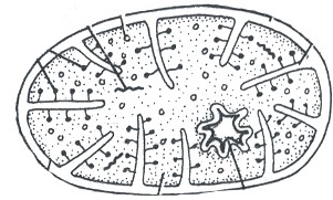
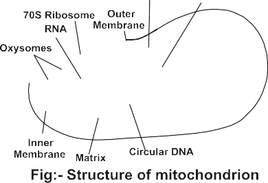
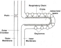
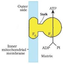
•The inner membrane and cristae bear electron transport chain and particles called Oxysomes or Elementary particles, F0–F1 particles or ETP (Electron transport particles) orATPsynthase
•The matrix also possesses single circular DNA molecule, a few RNA molecules, ribosomes (70S) and the components required for the synthesis of proteins.
•Mitochondrion is considered as semi autonomous cell organelle due to presence of DNA(rich in G–C ratio), RNA, 70S ribosomes and proteins synthesis systems.
•Themitochondriadividebyfission.
.
Functions of mitochondria
(i)Itisthesiteofaerobicrespiration.MostoftheATPareproducedbymitochondriaduringrespiration. Thus mitochondrion is called power house of cell.
Chondrioid.Thus Mesosome and Mitochondria are analogous organelles.
(vi)The gene for male sterility in maize plants is found in mt DNA. Thus it helps in cytoplasmic inheritance.
.
Endomembranous System
•Many membranous cell organelles are co-ordinated in their functions like ER, GB, Lysosome and vacuole so they are considered together as endo membranous system.
•Since the functions of the mitochondria, chloroplast and peroxisomes are not coordinated with the above components, these are not considered as part of the endomembrane system.
(iii)Tubules:Theyaretubelikeextensionsthatconnectcisternaeandvesicles.Diameterofeach tubule is 50–100 nm.
.
.
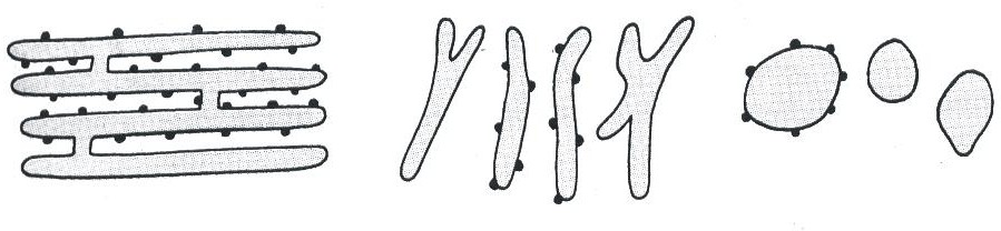
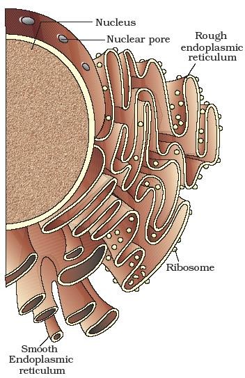
Types of Endoplasmic reticulum
On the basis of nature of its membranes, endoplasmic reticulum is of two types.
(i)RER – Rough Endoplasmic Reticulum
(ii)SER–SmoothEndoplasmicReticulum
.
•The ER often shows ribosomes attached to their outer surface. The endoplasmic reticulun bearing ribosomes on their surface is called rough endoplasmic reticulum (RER).
(vi)RER provides site for the protein synthesis and secretion, because it has ribosomes on its suface, Hence RER is frequently observed in the cells actively involved in protein synthesis and secretion.
(vii)SERsynthesizeslipids(phospholipids,chlolesterol),sterolsandsteroidhormones,visual pigments from vitamin A in retinal cells, Glycogen.
Special Points of ER:
(1)Microsomes: They are fragments of RER that are obtained by high speed centrifugation and Fragementation of cell.
(2)Sarcoplasmic Reticulum (S.R.): SER that occur in skeletal and cardiacmuscles are calledSR. It strores Ca++for release during muscles contraction.
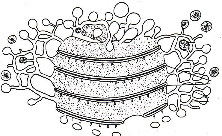
•Golgi complex is also Known as Dictyosome (plant golgi body), Lipochondria(Rich in lipids), traffic police of cell, Idiosome, Baker’s body, Dalton complex, Golgisome, export house/middle man of cell.
•Aplantcellhas10 –20dictyosomes.
Origin:
•GolgibodiesmainlyarisefromSER.
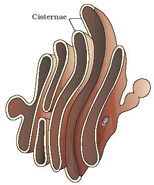
Structure of Golgi complex
•TheshapeandsizeofGolgicomplexdependuponthephysiologicalstateofthecells. Structurally Golgi complex is composed of four parts
2.Tubules:TheyformacomplexnetworktowardstheperipheryandtransfaceoftheGolgi apparatus. They interconnect the different cisternae.
.
.
.
.
.
.
.
.
.
.
.
.
.
.
.
.
Fig.StructureofGolgiapparatus Fig.Golgiapparatus
3.Vesicles: They are small sacs that arise from tubules. They are of two types smooth and coated. Out of them smooth vesicles contain secretory substances hence these are called secretory vesicles.
4.Golgian vacuoles: These are expensions of cisternae at trans face. Some of them act as lysosomes.
Functions:
(i)Secretion:
•Thegolgiapparatus principallyperformsthefunctionof packagingmaterials, to bedelivered either to the intra-cellular targets or secreted outside the cell.
•Golgi complex is a centre of reception, finishing, packaging and secreting for a varietyof materials in the cells.
•Aftermodificationsmaterialsarepackedinvesicles,thelatterarebuddedofffrommaturingfaceof Golgi body and released out side the cell that is called Exocytosis or revevrse pinocytosis.
(ii)Formation of new cell wall: Pectic compounds of middle lamella and various polysaccharides of the cell wall are secreted by Golgi complex.
(iii)Glycosidation and Glycosylation:
•A number of proteins synthesised by ribosomes on the endoplasmic reticulum are modified in the cisternae of the golgi apparatus before they are released from its trans face.
Note: Materials to be packaged in the form of vesicles from the ER fuse with the cis face of the golgi apparatus and move towards the maturing face. This explains, why the golgi apparatus remains in close association with the endoplasmic reticulum.
5.Lysosomes: (Suicidal bags or recycling centres or scavenger of cell)
•These are membrane bound vesicular structures formed by the process of packaging in the golgiapparatus.
•Theisolatedlysosomalvesicleshavebeenfoundtobevery richinalmostalltypesofhydrolytic enzymes (hydrolases – lipases, proteases, carbohydrases) optimally active at the acidic pH.
•The Lysosomes are absent in prokaryotic cells, RBCs, higher plants. They are abundently found in phagocytic and secreted cells like WBC, Kupffer’s cells, histocytes, pancreatic cells, liver cells.
•In plants, the tonoplast facilitates the transport of a number of ions and other materials against concentration gradients into the vacuole, hence their concentration is significantly higher in the vacuole than in the cytoplasm.
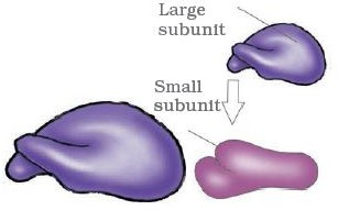
•Sizeanddensity oftheribosomesdependsuponsedimentationcoefficientintheultracentrifuge. It is mesured in Svedberg units (S).
•The two subunits of 80S ribosomes are 60S and 40S and the two subunits of 70S ribosomes are 50S and 30S. 0.001M Mg++concentration is requiredfor the association of two subunits as aresult intact ribosome is formed.
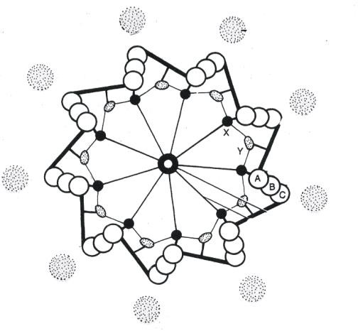
•The clear cytoplasm (Zone of exclusion) around centriole is called centrosphere or kinoplasmor cytocentrum. Both centrioles are commonly called Diplosomes.
•Centriolesareabsentinhigherplants.Althoughcentrioleisfoundinthoseplantsthatbearflagellate stage in the life cycle. e.g. Many green algae, Bryophytes, pteridophytes, cycads.
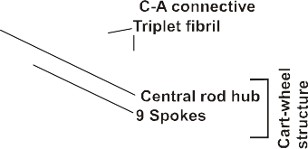
•Boththecentriolesinacentrosomelieperpendiculartoeachotherinwhicheachhasan organisation like the cartwheel.
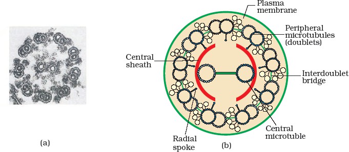
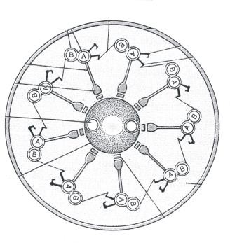
•Each centriole is composed of 9 peripheraltriplet fibrils of microtubules but in the central part these are absent. Thus centriole has 9 + 0 arrangement of tubules.
•The size of each peripheral triplet fibril is 25nmand It consists of threesubfibrils– C, B,A fromoutside towards innerside.
•Alinkerconnectstwoperipheraltripletfibrils insuchawaythatAsubfibrilofaperipheraltripletfibril is connected with C sub fibril of adjacent peripheral triplet fibril. This linker is called C– A linker.
•The main function of centriole is locomotion and the role of centriole in cell division is secondary function.
•Centriole is surrounded by amorphous structures called massules or perecentriolar satellite. Massules act as nucleating centre for the growth of microtubules during Aster formation. Occurs in S-phase.
•Thus new centriole arises from pre-existing centriole in S phase without presence of DNA due tomassulesCentrioles take parts in synthesis of Basal bodies, cilia, flagella, spindle poles.
•The central tubules are connected by bridges and is also enclosed by a central sheath, which is connected to one of the tubules of each peripheral doublets by a radial spoke. Thus, there are nine radial spokes.
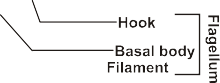
•A subfibril has two side arms or lateral arms composed of dynein protein. Out of them outer arm has hook. Inner arm show ATPase activity. It also generates force for the movement of cilia thusit is considered as locomotory motor for cilia.
•Theywere observed from endosperm of germinating castor beanseeds. These are commonin Neurospora. Germinating oily seeds of castor, groundnut and cucumbers, Yeast.
•J. Hammerling proved the role of nucleus in heredity, growth and morphogenesis. He proved that nucleus is the master organelle of the cell.
•Interphase stage is a best stage to study about the nucleus due to having highly extended and elaborated nucleoprotein complex known as chromatin, nuclear matrix and nucleoli (One or more spherical bodies).
•Multinucleated condition is found in some organisms this condition is called syncytium (arises due to fusion of cells) E.g. Ascaris or coenocytic (due to repeated nuclear divisons withoutcytokinesis) E.g.Vaucheria, Rhizopus.
.
Shapeandsize:Thesizeofnucleusis5–25..
•Thesizeofthenucleusdependsonthevolumeofcell,amountofDNAproteinandmetabolic activity of cell.
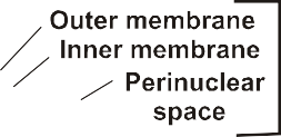
•The perinuclear space forms a barrier between the materials present inside the nucleus and that of the cytoplasm.
•The outer membrane usually remains continuous with the endoplasmic reticulum and also bearsribosomes on it.
•Atanumberofplacesthenuclear envelopeis interruptedbyminutepores, whichareformedbythe fusion of its two membranes.
•ThesenuclearporesarethepassagesthroughwhichmovementofRNAandproteinmolecules takes place in both directions between the nucleus and the cytoplasm.
(2)Nucleoplasm (Karyolymph): The nuclear matrix or the nucleoplasm contains nucleolus and chromatin. It is jelly like fluid, its pH is 7·4 ± 0·2. It is reservoir of nucleosides, enzyme of DNA and RNA synthesis.
•Itislargestpartofnucleus(35%)anditisdense,DNAfreesubcellularstructureandcontentof nucleolus is continuous with rest of the nucleoplasm is it is not membrane bound structure.
•Usually 1–4 nucleoli, are found in a nucleus of diploid cell. At least one nucleolus is found. 1600nucleoli have been reported in the oocytes of xenopus (An amphibian).
•NucleolusisasiteforactiveribosomalRNAsynthesis.Largerandmorenumerousnucleoliare present in cells actively carrying out protein synthesis.
•Plants generally have larger chromsomes than animals and amongst plants, monocots have bigger chromosomes than dicots.
•A single human cell has approximatelytwo metre long thread of DNA distributed among its fortysix (twenty three pairs) chromosomes.
•The interphase nucleus has a loose and indistinct network of nucleoprotein fibres called chromatin (contains DNA, histones protein, nonhistones protein and RNA).
•Duringcelldivision,dehydrationandcondensationofchromatinnetworktakeplaceasaresult chromatin is converted into chromosomes.
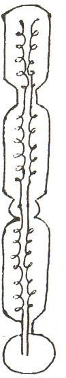
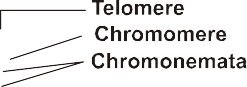
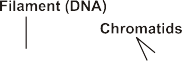
(iii)Chromonema:Thereistwochromonematawhenchromosomehastwochromatids. Chromonemata is coiled structure.
.
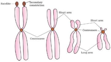
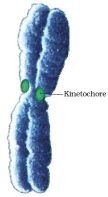
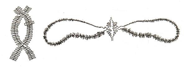
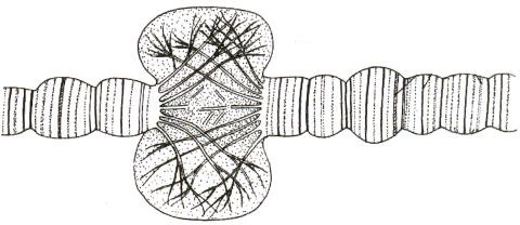
(iv)Centromere (Primary constriction): Narrow non stainable area where two chromatids are joined.The surface has disc or kinetochore on either side for attachment of microtubules belonging to chromosomal fibre.
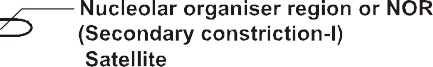
(v)Secondary constriction: They are narrow areas of two types. NOR and joints. NOR or nucleolar organiser region is secondary constriction-I capable of forming nucleolus in telophase. It is found on chromosome number 13, 14, 15, 21, 22. Joints or secondary constriction-II are areas involved in breaking and fusion of chromosome segments.
(vi)Sometimesafewchromosomeshavenon-stainingsecondaryconstrictionsataconstant location. This gives the appearance of a small fragment called the satellite.
(vii)Satellite: It is knob like part distal to NOR. The chromosome that bears satellite is called SAT (Sine acid Thymonucleinico) chromosome.
(viii)Telomeres: These are nonsticky terminal ends of chomosome or seal ends of chromosomes. They prevent the sticking of one chromosome with other. They are rich in guanine base. Telomerase enzyme is (required for replication of) this part of chromosome.
Special type of chromosomes
.
S.No.
Lampbrushchromosomes
Polytenechromosomes
1.
TheyfirstlyobservedbyFlemming.
TheydiscoveredbyBalbiani.
2.
Thesearefoundinyolkrichprimaryoocytesof Amphibians like Newt (Triturus), spermatocytes of many animals, giant nucleus of Acetabularia.
Theywere observedinthecellof salivary glandsofChironomuslarvaofDipterian insect. These are also found in malpighian tubules, endosperm, antipodal cells and salivary glands of Drosophila.
3.
Theyarefoundinpermanentdiplotenestage of meiosis.
Theyarefoundinpermanentprophase stage.
4.
Thesizeupto5.9mm(5900m).
Thesizeofpolytenechromosomes is
2000m.
5.
Special Characteristic: The axis of lamp-brush chromosome is composed of DNA and matrix ofRNAandproteinsItslateralloopshelpin synthesis of RNA and yolk.
Special Characteristic: They become giant due to endomitosis or endoduplication. Large swellings are foundonsomeplacesofeachstrandthat arecalledpuffs(Balbianirings).Inpuffs DNA is uncoiled for rapid transcription of RNA.
.
.
.
.
.
.
.
Chromosomalpuffor Balbiani ring
.
.
Dark
KnobBands
.
.
.
.
Interband
.
.
.
Fig:-Structureofpolytenechromosome
.
Karyotype:
Chromosomeshavesomespecificfeatures
(a)Numberofchromosomes
(b)Relativesize
(c)Positionofcentromere
(d)Lengthofarm
(e)Secondaryconstriction
(f)Satellites.
All such features by which a particular set of chromosomes (chromosomal complement) can be identified, is called karyotype of a species or it is chromosomal complement of organism providing description of various aspects of all the chormosomes like number, relative size, position of centromere, length of arms and centromeric ratio, secondary constriction and satellites.
Idiogram:
A diagrammatic representation of karyotype of a species showing morphological chraracteristics of the chromosome is called idiogram.
(a)Lignin: It is formed by polymerisation and dehydrogenation of aldehydes and alcohols of coniferyl and coumaryl. It reduces the water content of the wall matrix and increases its hardness. The deposition of lignin on the cell wall is called lignification that provides strengthening to the cell wall.
(b)Suberin: It is fatty substance that makes the wall impermeable. It reduces the transpiration rate in plants. It is found in the cork and casparian strips of endodermal cells. The deposition of suberin is called suberisation.
(c)Cutin: It lies as a distinct layer on the outside of the epidermal cell wall. It is fatty substance that reduces the rate or epidermal or surface transpiration. Other substances may also be deposited in the cell wall such as silica (E.g.grasses), minerals waxes, tannins, resins, gums.
(9)Spectrin,ahelicalextrinsicproteinusedtoattachwithintrinsicproteinattheinnersideof membrane andmake cytoskeleton with microtubule andmicrofilament
(30)Protoplast: It includes all the livingconstituents of the protoplasm.Nakedcell without cell wall is also called protoplast.
(31)Calmodulin: Calcium protein complex often associated with microtubules and microfilaments, taking part in motility and regulation of certain enzyme systems.